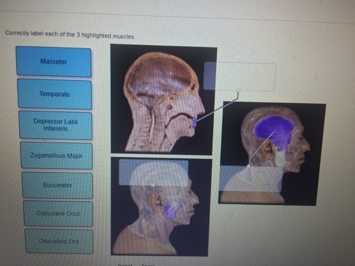
Correctly label each of the 3 highlighted muscles. – Correctly labeling muscles is crucial for understanding their anatomy, function, and clinical applications. This guide provides a step-by-step approach to identifying and labeling muscles, along with an exploration of muscle types, structure, function, and assessment.
The complexities of muscle anatomy are unveiled, including the different types of muscle fibers, the role of connective tissue, and the mechanisms of muscle contraction. The practical implications of muscle labeling are also discussed, highlighting its importance in clinical practice and the diagnosis and management of muscle injuries and disorders.
Muscle Identification and Labeling
Correctly labeling muscles is crucial for accurate anatomical descriptions, surgical procedures, and clinical diagnosis. It provides a standardized language for healthcare professionals to communicate and ensures precision in medical documentation.
Step-by-Step Guide to Identifying and Labeling Muscles:
- Locate the origin and insertion points of the muscle.
- Identify the muscle’s action by observing its movement.
- Use anatomical landmarks and muscle attachments to identify the muscle.
- Refer to anatomical atlases and textbooks for confirmation.
- Label the muscle using the correct anatomical terminology.
Muscle Anatomy: Correctly Label Each Of The 3 Highlighted Muscles.

Muscles are classified into three types based on their histological characteristics and function:
- Skeletal muscle:Attached to bones, responsible for voluntary movement.
- Smooth muscle:Found in internal organs, responsible for involuntary movements.
- Cardiac muscle:Found only in the heart, responsible for pumping blood.
Structure and Components of a Muscle Fiber:
- Myofibrils:Contractile units composed of actin and myosin filaments.
- Sarcomeres:Repeating units of myofibrils, the basic contractile units of muscle.
- Sarcolemma:Cell membrane of the muscle fiber.
- Sarcoplasmic reticulum:Stores calcium ions necessary for muscle contraction.
Role of Connective Tissue in Muscle Function:
- Endomysium:Surrounds individual muscle fibers.
- Perimysium:Surrounds bundles of muscle fibers (fascicles).
- Epimysium:Surrounds the entire muscle.
Connective tissue provides structural support, transmits force, and facilitates nutrient exchange.
Muscle Function

Muscle contraction occurs through a complex interplay of calcium ions, actin, and myosin filaments.
Types of Muscle Contractions:
- Isotonic:Muscle changes length while maintaining constant tension.
- Isometric:Muscle generates tension without changing length.
- Eccentric:Muscle lengthens while generating tension.
Factors Affecting Muscle Strength and Endurance:
- Muscle size and fiber type:Larger muscles and more fast-twitch fibers enhance strength.
- Neural factors:Recruitment of more motor units and firing rate increase strength.
- Hormonal factors:Testosterone and growth hormone promote muscle growth and strength.
- Training and nutrition:Resistance training and adequate protein intake enhance muscle function.
Clinical Applications

Examples of Muscle Labeling in Clinical Practice:
- Surgical procedures:Accurately identifying muscles is essential for safe and effective surgeries.
- Physical therapy:Muscle labeling guides rehabilitation exercises and interventions.
- Imaging studies:Labeling muscles on MRI and CT scans helps in diagnosis and treatment planning.
Assessment and Evaluation of Muscle Function:
- Manual muscle testing:Assesses muscle strength against resistance.
- Electromyography:Records electrical activity of muscles during contraction.
- Biopsy:Examines muscle tissue under a microscope to diagnose muscle disorders.
Implications of Muscle Injuries and Disorders:
- Strains and tears:Result from overstretching or tearing of muscle fibers.
- Muscle dystrophy:Inherited disorders characterized by progressive muscle weakness.
- Myositis:Inflammation of muscle tissue.
Popular Questions
Why is it important to correctly label muscles?
Correctly labeling muscles is important for clear communication among healthcare professionals, accurate documentation of muscle-related findings, and effective diagnosis and treatment of muscle injuries and disorders.
What are the different types of muscles?
There are three main types of muscles: skeletal muscle, smooth muscle, and cardiac muscle. Skeletal muscle is attached to bones and responsible for voluntary movement. Smooth muscle is found in the walls of organs and blood vessels and controls involuntary functions.
Cardiac muscle is found only in the heart and responsible for pumping blood.
How do muscles contract?
Muscles contract when a nerve impulse triggers the release of calcium ions from the sarcoplasmic reticulum. Calcium ions bind to receptors on the surface of myofilaments, causing them to slide past each other and shorten the muscle.